

¶ Intro / Opening
Worldwide, cardiovascular disease affects the lives of hundreds of millions. Dedicated cardionerds everywhere are working hard to fight this global epidemic. These are their stories.
¶ Introduction to Cardiac Sarcoidosis Imaging
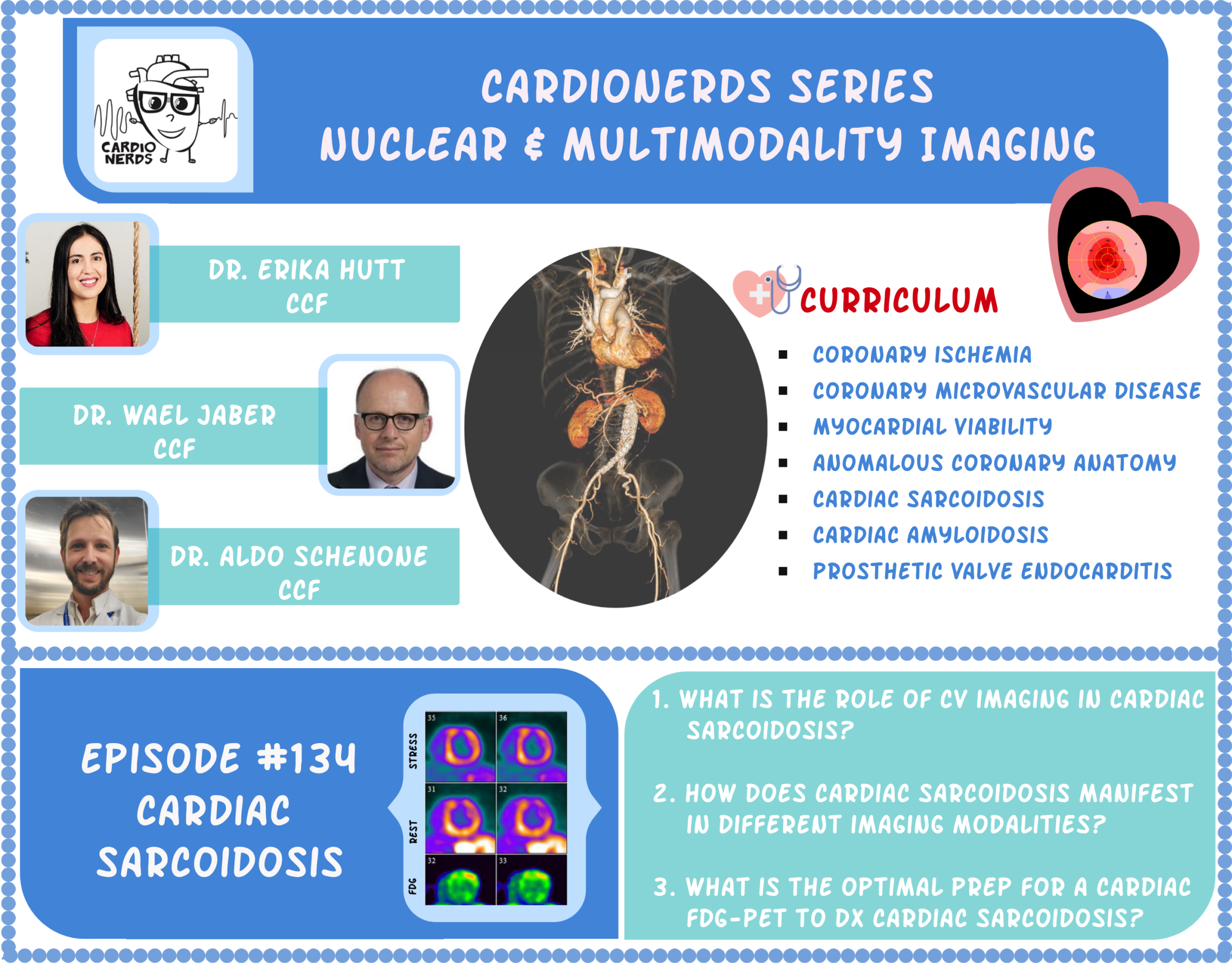
Hey hey, cardio nerds, it's Amit Coyle. Really excited to have you back for the seven-episode nuclear and complementary multimodality imaging series with Cleveland Clinic Imaging Expert Dr. Wilde Jebber. Future Cardiovascular Imager Dr. Erica Hutt as well as Brigham Imaging Fellow Doctor Eldo Scannoni. Make sure you check out episodes 99, 101, 102, 104, and 109.
When we discuss the multimodality imaging for coronary ischemia, coronary microvascular disease, myocardial viability, anomalous coronary arteries on myocardial bridges, as well as cardiac amyloidosis. Today we get to dive in to the multimodality imaging evaluation for cardiac sarcoidosis. As you enjoyed this discussion, be sure to refer back to episode 133, the last episode, where we got to discuss a fascinating case of cardiac sarcoidosis with our colleagues from the University of Chicago.
And to make sure you get every ounce of teaching from this incredible discussion, make sure you check out Incredible Notes on the Cardi Nerts website by Academy Fellow Dr. Hossein Khalid. Friends, we thank you for subscribing to and supporting the Cardi Nerds. This podcast is not meant to be used.
Used for medical advice, the views expressed here do not necessarily reflect the opinions or policies of our employers. Speaker disclosures are available in the episode description. There is no commercial or in-kind support for this activity. And make sure you claim free CME credit using the link in the episode description. And now, let's do some imaging.
We now come to our next patient. Tiresius is a prophet who lives in the underworld. That's actually where he met Odysseus. While showing Odysseus how to get back to Ithaca, Teresia suddenly collapses without a prodrome and later awakens feeling fatigued and confused. He was subsequently brought in by a cardiurence ambulance to the ED, where EKG showed normal sinus rhythm with a two to one AV block, a left bundle branch block, and frequent PVCs.
Naturally, he was admitted for a single P evaluation, and telemetry that night showed multiple runs of non sustained ventricular tachycardia with intermittent complete heart block. He was taken to the CICU by a diligent fellow who promptly threw in a transvenous pacer. Chest x ray shows a proper lead placement with no signs of pneumothorax, but it did show peculiar bilateral hyalur fullness. Now, as a young man, Therese is totally baffled.
He's been healthy all his life save for the occasional unexplained painful bumps on his legs. The fellow reassures him that he's in good hands, and the EP team would likely place a permanent pacemaker the very next day. Notably, he's not been exposed to AV nodal blockers, and electrolytes, serum lime antibody, and ACE levels are all negative. So Hefe, what's going on with him? And does nuclear imaging play a role?
¶ Initial Cardiac Evaluation and CAD Exclusion
So this is a uh a healthy person presenting with syncope, the uh sudden syncope we recovered from. The the things that go into your mind is this cardiac or non cardiac, but this is a a cardiac podcast, so we'll stick with the cardiac side of it and uh we'll go from So be before we we start.
uh doing a lot of fancy testing, let's do the the normal thing. So you're looking at the EKG, you describe a two to one A V block, you describe the frequent arrhythmia arrhythmia in this person, left ponder branch block. So this person who's young with no prior history of any uh other issue has evidence of conduction distance disease. So this is infrahistian disease. This is not normal as unless you're dealing with ischemic heart disease.
in the general population, much older age that can present too, but this person is active and healthy, so it shouldn't be an issue. So we start thinking beyond ischemic heart. So the first test of course the best tool we have in echardiology is echocardiography because it's non invasive, it gives us an idea about what's happening with the structures of the heart, the pumps, the valves and all these things, the pericardium, the water.
So I would do just first a screening echo. And a screening echo on this person is gonna show you either a normal function, everything is fine, or it's gonna show you some areas of patchy wall motion abnormalities that are not or are consistent with CA.
It's gonna be a little bit difficult here because of the left mandorbranch block. They're pacing now, you're pacing this person. So it's gonna be very hard to expect a normal synchronous contraction, contraction of the left ventricle and the right ventricle. So you're gonna be a little bit of the synchrony because of that.
But be it as it may, when we did the echo on this person here, we found that he has wall motion rheumatis with wall thinning in the basal septum and in the basal and mid inferolateral wall and anterolateral wall. Even with that, with what we call characteristic or pathogneumonic or very consistent, whichever emphasis point you wanna uh hit, consistent with potentially sarcoid if you tie it with the hyler filling on the chest x-ray.
Uh I think it is very imperative that we rule out C A D in these papers. Now given that your patient is young, you can do it the easy way, you can do it the traditional way. The easy way is to send this person for a a CTA. You can do a CTA of the coronary arteries if the coronary arteries are open and happy. Down now we're dealing with an inflammatory process with resultant scar.
If the CTA is showing us obstructive C A D, then we have to go down that path. Now there is a bit of overlap with humans and we can have multiple diseases or multiple things threatening our life, but it's rare. So I will start with a CTA. If you don't have a CTA or you're not comfortable with CTA or the age group is not conducive to do a CTA because of classifications and all these things.
you can go for a corner angiogram. But it's it's very important to start with that step before you start going down the fancy part of doing uh PET scanning or MRI or any of these other things because there is again a lot of overlap and you can miss CAD at your peril and the peril of the patient.
¶ PET and MRI: Complementary Tools
So this is what I think about this. I I start with an anatomic test to make sure there is start with an echo of course, then go for an anatomic test to rule out obstructive C A D. And from that point on you can choose an MRI to look for scarring or inflammation. Or you can do the pet pathway and look for scarring and inflammation both in the heart and extra cardiac, in the chest, in the lungs, and the liver. spleen, you know, guts and all the
You know, I I I think that people always try to put these things against each other. Should we do a pet first, should we do an M R should which one is better than the other? I think in a in a way they're complementary. M R I tends to be very good test initially, but I don't think it's Very good test to keep repeating.
Uh PET if you're planning on therapy is one of the probably the coolest tests we have because if you find inflammation you can follow that inflammation and follow if whether you suppressed it or not with serial pets, especially if you decide to go on and put a pacemaker and I C D and all these things. People talk about now we can do
MRI with all these things on, you can do them, yes, but your diagnostic yield becomes uh challenged with more uh hardware you put in. So in this patient we did a CTA and the CTA was completely normal, right? No coronary disease. And then now we we sent the patient because this is the toolbox I have, which is a pet. We decide to send the patient this patient for a pet.
¶ Cardiac FDG-PET Patient Preparation
Now although you're at now at a different institution than ours, so how do you guys do the pets on these patients and just walk us through the initial pet? This is not the follow-up pet, this is the initial pet on the Absolutely. It's key to understand how to prepare the patient for the bad F D G for for psycho, what we call the psycho protocol. We should start a little bit of a little bit of physiology and and why are we doing this thing?
So when when you're imaging the HARP with Ft G, which is a glucose analog, so although the HARP primarily functions with fatty acid, it uptakes a little bit of glucose as well. Especially those areas that are either ischemic or hibernating. So that's why it's so important to rule out uh any obstructive disease before embarking in this particular test.
So the second challenge is that we want the FTD to primarily be taken out by potential inflammatory cells within potential granulomas. So how we can make the heart not take any glucose and and that's where the preparation is key. So what we do here at the pregams is that we advocate for a prolonged fasting, you know, around ten to twelve hours. And that prolonged fasting is preceded by two meals that are hot in fat and proteins and has no carbohydrates at all.
And the whole idea is that you're trying to switch the metabolism because there's no ability or there's no offer of glucose at all. So the heart would switch the metabolism to purely fat. So by the time you inject the patient with the S Dg, so the heart would not or the mock car would not pick it up or hopefully would not be picking it up any any F Dg because we'll be f using fat as a fuel.
So it would only be the potential inflammatory cells within the granuloma that are gonna be there picking it up and you can see it with the image. So that preparation I think is key. It's important that the medication specialist is in the unit, is not taking anything by vein that has any uh dextrose. So we need to be really careful uh about that.
And making sure that any of the food that is the patient got the before the fasting has not in the glucose so so we can get a nice preparation. Because the worst case scenario if you don't do that properly, what you can end up having is having
some uptake in the myocardium that you don't know if is is actually related to the inflammatory cells or it's just normal myocardial uptake or what we call non specific. So I think that's some of the the key points in the preparation. So is pretty standard we pair rest in profusion along with the F T G imaging and and we try to avoid at all cost doing stress imaging. at the same time or at the same time or the same encounter as the
the SARCOD imaging because we don't want to induce ischemia that could potentially induce a FTG uptake by the myocardium. So those are kind of in big concepts. what we do. Uh I know the other centers use uh heparin prior to the scanning and and the whole idea is the heparin induced like policies and the concept is that if you give that just prior to the scan, you increase the offer of fatty acids to the myocardium, you're gonna again
reemphasize or keep pushing that fatty metabolism to the myocardium. And I know at the at the clinic where we're doing d that when I was there and then we were getting kind of good result. Here at the pregram, even without that, we're we're getting good images. That's very helpful, Aldo. So you know, we go through all this prep with a ketogenic diet, perhaps intravenous heparin right before the study to suppress myocardial F D G uptake.
So that whatever F D G uptake we see on the pet is reflecting uptake from white blood cells, right? That would reflect inflammation. And of course, you know, there might be some other non sarcoid, non-granulomerous inflammation causes of white blood cells, like perhaps you know, like a different kind of myocarditis. Another situation we had uh previously on the show was a wonderful case from Northwestern where they you know, they had a patient with a EF of forty percent coming in with VT Storm.
And they thought this patient may have sarco, so they did a sarcoid PEP, and the entire myocardium diffusely lit up, so they thought, oh, maybe this was an inadequate prep.
And so then they repeated the study and the entire myocardium lit up again. Then they did a cardiac MRI I believe and it showed that yeah there actually is inflammation. And subsequently because of family history and the overall picture, Um, sent a patient for genetic testing, and it turned out to be uh DSP Daphne plaque and cardiomyopathy, which is a left dominant errythmogenic cardiomyopathy, and in a small cohort of patients.
In a series earlier this year, that can present with these like acute bouts of cardiac injury with an inflammatory picture, a troponin leak, chest pain that all for all the world looks like ACS. And then also I think immediately post MI if you do an FTG pet, then you know you'll just have uh inflammatory uptake from the post MI situation and not necessarily reflect sarcoid uptake.
But when we do these the sarcoid F D G PET, we also do it with a resting profusion PET scan, rubidium or ammonia. So Hefe, what information does that add to the F D G PET in diagnosing the sarcoid? So the most important point I think is uh although hit on those on some of these points which is the prep of the patient. So first rule out coronary artery disease and then the preparation of the patient. I cannot
talk enough. We can spend two hours talking about how important is preparation of the patient. If you do these patients without prep, you're gonna end up with a very confusing test. So you know I just have to say one of my pearls of wisdom, if you wanna say that So years ago when uh Stalin was uh ruling Russia or the Soviet Union, so there was a very famous uh Russian poet and that Russian poet Akmatova
She's famous. He loved her poetry. He loved everything about her. And she was always she was a dissident but she was careful about not criticizing him. But she's always, you know, saying wonderful things about communism and all these things. And one day, you know, he put her on house arrest and made her life very miserable. So his advisors went to him and told him, you know, why would you do that? You know, she loves she loves you. She loves the Soviet Union. She's doing everything, you know.
So he said it's not important for you to love the Soviet Union. It's important for the Soviet Union to love you back. And that's the same thing here when we talk about pets. You love pet, but the pet has to love you back. And it has to love you back in a way where you have to know how to approach this test with first some humility about its limitations, which you touched on.
One is inflammation is universal, so we don't know we cannot tell one inflammation type from another. Two, the prep is extremely important. Now Erica, I know you uh we're gonna answer your question, uh Amit in a second about about the pet specificity and all this stuff. But Erica, you you recently published a a study that you've done with the with Paul Kramer here about how to prep these patients.
Can you just tell us a little bit about it just briefly so we can set the stage of how we use it here? Of course, Doctor Javer, thank you. And actually Aldo's description of PrEP looks very similar to I think what we used to do, but we're trying to transition to this new dietary preparation that Dr. Kramer is actually trying to standardize that our institution. So what we found is that many patients actually fail to suppress myocardium.
with the standard preparation they used so that fasting for twelve hours and the low carbohydrate, high fatty acid diet was not really well completed by the patient. So we know that the compliance is not a hundred percent. And that is why he tried to standardize it in this other manner, which is with the use of a ketone shape.
So it's basically a baby formula, like Abbott's baby formula based on ketones. And he started doing it on patients, mainly inpatient,'cause there was more control over those patients. So giving the ketone formula, the breakfast before the study, lunch before the study and after lunch, NPO for the study the following day.
And we've actually seen like on significant amount of patients that had failure to suppress myocardium with just a standardized diet, that was with this new protocol, we've been able to adequately suppress Glucose metabolism in the myocardium and see the inflammation. So I think it's it's something that we're working on reporting in a higher volume of patients to see if it's
It's obviously not just random luck that we're seeing this or if it's actually working. So hopefully we'll be publishing something pretty soon. So Erica, that that that's very interesting and definitely anything that we can do to improve the preparation prior to a pet sarquid is is
is gonna be always well received. I think at one point that uh I often see in clinical practice is those patients that are in the unit that either they're intubated or an NG tube. And yes, certainly I think the most important part of the preparation is this prolonged fasting. But we often get asked how we can feed these patients, you know, high fat. high protein perhaps kind of meals. And what we have done here is just through the end tube we
We have provided them with some sort of oil preparation, olive oil, and and you can you know provide them with that as a change of for a potential meal. And we follow that with the prolonged fasting. I I think the other important concept is in going back to you guys new preparation or or proposal is that it's often a challenge for for patients that uh are vegetarian or vegan then in terms of how you
make them follow the diet, given that, you know, the the mainstream in terms of this diet preparation is is meat. It's it's easy for someone who eats meat to follow them, but you know, for for people that prefer to uh stay away from those is is hard and we often, you know, rely on either tofu and and and and oil. It's very challenging to come up with a kind of a good meal that can replace
kind of the classic meat and so forth. So I think it would be very interesting what you guys come up with and and maybe it's it's kind of a great solution for those who want to do a preparation without having get meat and some of these kind of protein and animal product
Agree, Aldo, this is extremely important. Then then we move to the question you asked earlier, which I digressed from uh because I wanna stress the importance of preparation. This is the test without a good preparation is a useless test. So it will not love you back. You will love the test, but the test will not love you back. So I I heard somebody say that a test without a good preparation is preparation to fail.
That's that's that's not only for a patient to fail and fail yourself and fail the patient and then you end up going down a path which is not actually it's not actually it's detrimental for the for the For the patient, because either you missed the diagnosis completely or you misdiagnose the patient with sarcoids and end up treating them with highly toxic medications, and then you end up with a big problem.
And you make it difficult for your colleagues if they're reading these tests after you in the future to to compare the tests safely. So you all these steps are extremely important, making the right diagnosis, making it easily, making it correct. And setting the stage for a proper follow-up and proper treatment regimen with proper follow-up with imaging. If you don't do it right the first time, I think you can end up with a very big problem. So preparation, preparation, preparation.
¶ Interpreting FDG-PET for Sarcoidosis
Now, we move to why do we do perfusion images on this spaceship. So again, because of the non-specific cardiac uptake of FTG, FTG is normally taken by the heart, even in normal situations, although we talk about fatty acid metabolism and all these things, you still have FTG uptake of the heart. So we start with a resting image to one, define any perfusion defects. So if you have perfusion defects, that's actually kind of a good news for the imager, not for the patient, but for the image.
So uh if we have no perfusion defects, everything is normal, that tells us that there is no scar almost in the heart. If you end up with situations like that, which we'll talk about in a second, that decreases the specificity of the test. Because now you're going to image with FTG after that. So you have a normal perfusion, complete anomaly perfusion. Now you're gonna see areas of Ftg uptake with normal perfusion. Now are we seeing inflammation? before we're seeing scar in this situation or
Are we seeing just a normal pattern of FDG uptake in the heart? Because the heart will take up FDG. And despite all our efforts to suppress that. by giving them the diet and all these things, we can still fail to suppress. And we can still fail to suppress not uniformly, we can fail to suppress locally. Let's say in the areas of papillary muscle and the areas of the basal septum and the areas where we think they're sarcoid typical.
So those are the difficult cases I think we face is when we have totally normal perfusion, but some patchy FDGR. So let's say we do a perfusion study and now we have, as we saw on the echo in this patient, basal septum has perfusion defect, the basal inferolateral wall and mid-inferolateral wall have a nice defect. So now we have defects on the perfusion images. Now we go to the FTG images and we have two scenarios if we're successful in suppressing the FTG.
We can have uh no uptake in those segments, Ftg uptake, and that will tell us that these areas are scarred because we have now a perfusion defect with a matching FTG defect. So these are areas of scar.
Now those patients can be prone to have arrhythmias, of course, can be prone to have especially if it's a basis septum in those areas to have conduction defects. So that's a typical what you wanna see and we'll be glad you saw it because now you're matching the whole history with the electric side with the uh imaging.
Or you can have a a more hopeful situation where you can see defects in the inferior septum, defects in the inferior lateral wall, and now you see enhanced FTG uptake in those segments. So you say, okay, good. Now we have areas of inflammation in the heart. And then fear acceptum and fear infrared.
So now your challenge is uh these are areas of inflammation, we're very happy. Now you saw also areas of inflammation, which is important, we didn't talk about that. Every time we do sarcoid imaging as an initial image at the Cleveland Clinic, with PET we do a whole body sarcoid imaging. To pick up extra cardiac sarcoid. Because if you have extra cardiac sarcoid and we have conduction of math, then we have cardiac involvement, even if we don't see it on him.
So in this patient we saw the hyaluronymph adenopathy light up. Sometimes we see a lot of mesenteric adenopathy that lights up. Sometimes we see things all over the body. So that's important also to to pick up. So we did the study, we did a rest perfusion image with rubidium, as we do it generally here. We have access to rubidium, but we do rubidium.
And then we do FTG, we saw inflammation and inferior infralateral wall and basal septum. Now the challenge is what to do with this patient because you have temporary wire pacing on this patient. Do you start treating them and hope for recovery of the conduction? Or do you go out and how long do you wait? We don't know.
You know, temporary wires cannot stay in for weeks. Or do you start just go ahead and because the patient had an event, you go ahead and just put a combination of a pacemaker and I C D. And then what's the ejection fraction and all these things? So with the with the respir fusion image also you can get the ejection fraction on this patient. You can get an idea about what's happening with the R V function on these patients. A lot of patients with pulmonary sarcoid will have
uh bad RVs too because of pulmonary hypertension and things like that. So you can see those things by PET in addition of echo also. So that's the gist of of why we do rest perfusion images. What kind of results you can see with the PET, you can see if you're successful in myocardial suppression, you can see either scarring or inflammation. And just a a quick clarification, so if I see a perfusion defect and a matching F D G uptake,
then I imagine that can mean one of two things. Either there's inflammation and there's also, you know, s already scar there. Or there's inflammation and because of the edema, microvascular compression, there's a profusion defect, but that territory is not necessarily dead and scarred and is salvageable. with uh anti inflammatory. Is that fair that there it can go two ways?
I would say that's a difficult question to answer. The difficult thing with PET is although it has a very good spatial resolution, but we cannot tell subendocardial from the rest of the myocardium. So just think about anything we see as a continuum. So you can be having leftover inflammation in tissue that's below the spatial resolution of what we can see by PEC.
You can have this combination things. Actually I just recently read a pet with with Erica just last Thursday where we said there is a combination of ischemia and hibernation in that territory because you can have both. You can have hibernation and uh scar in the same territory. Remember, these are islands of things and we're trying to be all or none and it's not it's not that easy.
So you can have islands of scar and islands of inflammation, you can have islands of normal tissue and islands of inflammation. Now if you see normal perfusion and inflammation, you can have that too. So you can have all these things, but we're trying to be here more binary in our thinking, which is not fair for for the uh normal human body to function in human body doesn't b function in binary things.
So that's why your question is important, but the easiest things if you refer to the to the original paper from Ron Blankstein about how to diagnose these entities. The easiest ones are if you have a matching defect, you have a perfusion defect and matching FDG defect, then that Scar. And the next easier one is if you have a defect by perfusion and active inflammation in the same areas of the perfusion defects, that's inflammation. And everything else is difficult to reach.
Now, the easiest one to read is if you have a normal perfusion and you have no uptake whatsoever in the myocardium. And that's easy too. That's very helpful. And like I think many yes and no questions in medicine, the best answer is that it's complicated.
¶ Addressing Ischemic Memory and ICU Patients
Thank you, Hefe, that's a great explanation. And you already explained this previously, but you have a patient that comes in with a complete heart block or uh V T and you want to rule out ischemic heart disease first. And in a perfect world we do that with a CTA or a coronary angiogram, but you know that many of these patients that come to the cardiac ICU have renal dysfunction and so then we're dealing with a situation where the physician wants to rot ischemia with uh a pet Stress rest.
And then also SARGORD given the high clinical suspicion. So as Aldo alluded before, we don't like doing those tests the same day because there could be ischemic memory and you could have FDG uptake in areas of resting perfusion defect. So how how do we go around that and is there a way we could actually answer both questions with pets?
Again, it's complicated. So that's why I always pray when I'm reading one of these pets that the patient had an atomic study before. Because that makes my life, your life, everybody's life. so if Stress and then F D G now.
It's important again, although talked about ischemic memory and stuff like that, it's important not to also do a treadmill stress test because a lot sometimes you get these patients and they do a treadmill stress test. Let's say you have an ammonia, access to ammonia, do a treadmill ammonia. Now what you have is you have the uh skeletal activity of the FTG, and you try to image these patients with FTG later, and now you all you see is the lighting up of the musculoskeletal system, not the heart.
So that you have to be careful of not doing also. These patients should not go and engage in a running marathon or or even any strenuous physical activity before you do F D G on them because that can be Also enhancing the small musculoskeletal system. So you preferred patients as a patient in the hospital with bed rest, getting the ketone shake that you give them, and then come down for there.
Now in general we don't do a stretch treadmill on these patients, we do or ragadinocent pet on these patients. So if they had a panatomic study, it's easy. If you see ischemia in these patients, you can attribute that to microvascular disease, to the inflammation. And we've seen we have many, many cases where we do rest stress and then FTG for sarcoid. where they have normal correct arteries and we've seen ischemia in the inferolateral wall, antrilateral wall.
in segments that can match circumflex or can match rca territory and that can be attributed to edema and inflammation in those areas and therefore you're not able to vasodilate properly with dracodenosine and then end up with an esophageal. Now, the difficult part if you use ischemia in these areas and then you can see FTG uptake in those areas. Is this what Aldo was talking about as ischemic memory? Now you can avoid doing that by staging the test.
You can do a rest F D G on day one and then you do the stress test on the day two. So you do the testing on day two. So that by then you you avoid that kind of uh mishmash things and stuff like that. Now you know I guess you're keeping the patient in the hospital and all these other things. But you can play a little bit around and and then stage your test in in two different days by doing the rest in F D G day one.
To avoid ischemic memory. So ischemic memory is basically when you induce ischemia, whether with drugodenosin or whether with exercise. And you go ahead and you do F D G afterwards, let's say half an hour or twenty minutes later or thirty minutes later, now you if you are
lucky and you have induced real ischemia, you have switched the metabolism from fatty acid to glucose, and you go to inject glucose, that switch is not quick. So it happens over let's say minutes or whatever, but it doesn't go away over minutes. It can persist. So you go to inject FTG, now you're picking up the ischemia or the switch from fatty acid to FTG that happened much earlier.
And that's what we call ischemic memory. This has been published and studied and talked about before and actually in in reading normal pets for ischemia it can be very reassuring. So if you end happen to do a patient with rest stress and FDG for ischemia, not for sarcoid, and you saw the areas that induced ischemia, you saw they have FDG, you know this is not an artifact.
You know, this is actually real. So it can be actually an insurance policy on reading the stress test. So the I like it sometimes when we do it that way because that's where we do it. uh it increases the certainty in what we call the ischemia. So it's not a lateral artifact from breathing or from reconstruction of the images or C T superimposition on the on the transmission images and the emission images and things like that.
So that's ischemic memory. But in general we we like to do rest and F D G alone and get away with His concept of ischemic memory increasing Ftg uptake is fascinating and I'm remembering Erica in an earlier discussion when you said that it's just so amazing that you can understand what's happening at the cellular, subcellular, mitochondrial level. with nuclear functional imaging. It's just it's really fascinating. So going back to our patient
Yeah, these patients with sarcoidosis, their manifestations, right, depend on the the distribution, patchiness, extent, severity of inflammation, scar, right? So you range in your clinical manifestations from heart block, ventricular arrhythmias, you can get atrial arrhythmias, heart failure, uh a multitude of causes of pulmonary hypertension, possibly with insulant cord pulmonal RV failure. These patients can get pretty sick, right? And so you may see them on the first encounter in the ICU.
And I'm remembering, again, we we talked about this a little bit in an earlier discussion, but our esteemed panel here, Erica U, Eldo, Dr. Jabert, wrote an article about the multitude utilities of nuclear imaging in ICU patients. So do you wanna talk a little bit about the use of nuclear imaging just in you know, how it's relevant in patients with conduction disease and arrhythmia that may land in the ICU?
course and this is actually a paper that is about to be published and Aldo is the leading a author on it. And as we've discussed previously, the advantages that nuclear imaging has over other imaging modalities like cardiac MRIs, the fact that there's no interference with
intracardiac devices. So a temporary pacemaker like in this patient wouldn't interfere with the image as it would with uh cardiac MRI. There's no need to have normal renal function or normal hepatic function, so there's no limitations. And the test is actually a very quick test. So even patients that are intubated or have devices like intraverted balloon pumps that we've discussed before can go to the nuclear lab safely with adequate transport, et cetera, and not have to be there forever.
If they have hemodynamic instability, you can take them out of the scanner pretty quickly, which is not the case with a cardiac MRI. So all those things I think favor nuclear imaging in the critically ill patient. However, we always try in a patient with cardiac sarcoidosis to get a cardiac MRI before putting in a device.
So if we happen to be lucky and the patient doesn't have any hemodynamic instability or any unstable arrhythmias, then I think at the clinic at least we we encourage getting that initial cardiac MRI that El Hefe just mentioned, that we we always like to have that initial test that is very helpful in them.
the units are becoming more complex nowadays and we're seeing very sick patients completely different to what's in the past of just a coronary unit. Now it's a really complex cardiac intestine unit. So nuclear imaging can help you in identify what's what's going on with the patient, either sarcoid or infective endocarditis or, you know, viability, potential ischemia, and select a group of patients that that you need that information to move on in care. So I think I think it's that's relevant.
¶ Clinical Triggers for Sarcoidosis Workup
So now that we've discussed the utility of sarcoid PET in the evaluation of cardiac sarcoidosis, Aldo, why don't you tell us about other imaging modalities that are useful to delineate cardiac sarcoidosis? Uh absolutely Erica. I think this is kind of an important topic. Before we get into how we use the different modalities, one question that always comes up is is when do I need to do this? When do I trigger uh a work up for patients with either sargoid or or suspicious for sargoid.
from a big overview, I think if you have a patient that has known extracardiac sarcoid and that patient present with either palpitaceous or arrhythmia or syncope or presyncopy has any new anormalities in the ECG or any new funding on the echocardiogram, that's already enough for you to to go ahead.
and interrogate if the patient has any cardiac involvement. The second group of patients is those that even if they don't have history of SARCWAD, they present with in a clinical scenario that is very concerning for SARCWAT. And we're talking about people who present with Hydrogree A B block as the patient that we're seeing here, and they have no other potential costs, especially those Jone Fox.
you know, especially those uh under the age of sixty, uh similar way those those patients presenting with ventricular tachycardia or Bt storm and and there's no clear explanation for for that, I think that's another scenario that is useful to interrogate the presence of of of SARCOL even if the patient doesn't have
uh history of extracardiac cirquid. And as we're gonna see at the end, in certain scenarios you would incline to do one versus the other modality, depending on what was the presentation of the patient. But you know, getting into the modalities we'll we'll zoom up uh at the end.
¶ Echocardiography and Cardiac MRI Findings
So I think I'll hef uh pointed out echocardiography is the foundation of imaging for us in cardiology and an easy test we can do at the bedside, easy, quick, no radiation. and and give us a glance of what's going on and you can you can identify findings that can be suggestive of sarcotin echocardiography but I think the the most important concept to understand here is that the echo is not sensitive nor specific. And in fact, even if you have a
A relatively normal echo, the negative predictive value of like 32%. So by no means you can say, oh no echo is normal, we're done. This patient doesn't have sarcoids. So but if you find uh abnormalities such as either low EF, uh we have this common pattern of either uh uh basal septal dyskinesis or thinning, which you often see in in these patients with sarco, especially those who present with uh you know a complete heart block or high degree block.
The other area that uh Sarko likes is the baseline ferrolateral, as I Hefe pointed out, and you can see the same similar pattern, like this kind of really weird bite. kind of thinning of the of the wall which is looks not coronary not coronary distribution, which is diarrhea thin in this kinetic, then uh you need to increase your suspicion that this is probably or could be a presentation of Sarquet.
There's some data out there in terms of strain. There's a lot of debate. There's some data suggesting that And you can use radial strain to discriminate SARC weight, but then there's some other data saying that it's not radial strain, but it's actually longitudinal and circumferential. I can help you in that. I think although there's that out there, I think you cannot hang your head on on on just straining.
It's a clinical integration with the imaging and using echo as a first step just to kind of lead you to the following imaging. You know, I think the the two big tasks. in in the assessment of potential cardiac sarcoid, as we has been uh discussing. First is PET with FDG, rest, perfusion and FD im imaging. On the other side, a cardiac MRI has proven to be a very powerful and very uh useful
As a rule of thumb and and kind of Eric already kind of pointed out a little bit about that, the strength of MR is it has a high negative predictive value. So in those folks that we have let's say extra cardiac sarquid, but you know, they have some palpitation, there's some suspicions, but you know, the presentation is not like slam dunk and having an MRI is important because if you have a completely normal MRI
Yeah, you know, that test provides you a, as I said, a high negative value for the presence of cardiac involvement. And and what do we look for in cardiac MRI? Well what we look is is for signs of infiltration. So MRI can provide you with, you know, again, all the visualization of the cardiac function, dyskinesis, akinesis, wall thinning, similar to what you get in Echo. But also provide you with the opportunity to look for potential myocardial edema, focal or diffuse in
Although we can do that, in all honesty, the the um T2 imaging, which is what we use for for edema along with other other sequences, is not necessarily the best. And when you compare uh uhcarial edema inflammation with MR compared to PET, PET is definitely a winner. But we have ways to look for edema and with T two imaging and and we're looking in in circle the presence of these
patchy focal areas of increased signal in in T2 weighted imaging that could suggest there's some areas of edema for inflammation. The strength of the MR is the capacity to identify areas of infiltration. and and we do that by uh using legal rhythm enhancement. So we inject uh the gallinium and we wait
you know, 10-15 meta to that gadolinium to really get into the myocardium. And then in the areas that you have either inflammation or scarring, you're gonna see that the myocardium is gonna take up more gadolinium, it's gonna it's gonna light up.
But that can happen with any inflammation or any fibrosis. How can we tell if if we're in the presence of potential sarcodoses? Well, so The the classic pattern of sarcodosis is is the presence of uh a very intense gallium optique which is patchy, there's focal, ideally multifocal, that inc will increase your kind of your certainty. But also you see that that area of the wall, instead of being thin out, is actually expanding.
If you think about it, when you have an infarct and you have fibrosis, you have replacement fibrosis. So what that means is the myocardium you lose the myocytes and that's replaced with collagen, and you replace the myocardium and the mycardium
things out. When you have an infiltration, you you basically put more stuff, you stuff the myocardium. And the myocardium even though have a lot of gadolinium, but gadolinium is going to the inflammation, to the scarring, to the inflammatory cells and everything. That actually doesn't necessarily replace but actually expand the martyrdom. So so the classic pattern kind of the textbook is that one that is
Is focal, involving multiple regions. And also you see that it expands the myocardium, and there's some degree of association with edema on T2 weighted imaging. The challenge is here is that.
¶ Complexity of MRI Interpretation and Modality Integration
Yeah, we all like to have that kind of classic pattern, but that doesn't happen happen often and and Sarquet is kind of the T V the tuberculosis that we have in medicine that can present us anything. Psychodosis is a TV of cardiology. So psychosis can present in any pattern. So you can have linear LGE, can have uh more diffuse or or or areas of i uh insertion point of the R V. So it can present with a variety of of LG, not actually the classic, you know, multifocal, expansive
kind of LGE. So so that really add to the complexity of interpreting carioxarcosis. So So that's why when we look at cardiac more, yes, it has a high negative relative value. If it's totally normal you are you know you can be reassured that most likely this patient has nothing. But if you have of course a very classic pattern, then you are highly suspicious.
of having circuit. But if you're in the middle, like you know, a little patchy here but not classic, then you still have a little some degree of uh uncertainty. And that's why when we look or we talk about advanced imaging, we talk and we say that the MR and PET FTG are actually complementary. It's not that the one is better than the other one. They look at different things and and then actually they're more powerful
when you uh use them together. For instance, if you see a uh a patient that you see these, you know, uh focalers of uh uh LGE which is a expansive a little bit and you do an an an F D G PED you see there's that's areas lighting up, but there are other areas as well that are lighting up with maybe some areas of perhaps you know perfusion defects and you see extra cardiac lighting up on pet.
then you know your your certainty of the diagnosis goes up. On the other a area where you see these areas of, you know, some L G E but then uh there there's no F D G on PET and there's no extra cardiac, then it's a little bit harder to kind of come up with a diagonal. The other challenge is that I mean, in the past we have talked about
stages of the disease and we traditionally has described like maybe you have inflammation initially, that inflammation can create a profusion defect from the inflammation itself, microvascular dysfunction and so forth. And maybe you transition into
you know, a little bit of inflammation in scar and then finally you have end stage have all scar out and you you can even get to that point where you have thinning of the wall because scar is is what predominates. But now we have seen that you know, rather than stages, this might be actually patterns of disease and and it's not necessarily that linear, although there's some linearity on the background, but for the most part you can
see patients that present with one pattern, you treat them and that pattern changes and then the the disease can come back in a different pattern and and is is adding to the complexity of the disease and our understanding from an imaging standpoint in there. So The way that we incorporate all this finding I think it's important to, you know, because as you can see it's very complex and and and looking at the image and interpreting the images but also is even more complicated to report it.
to the clinician. And I think going back to what what Er Hefe said, I think the initial paper by Ron and and which kind of tried to get at this and try to determine what the the likelihood of disease based on patterns and it that's a good way to talk and to have a common language. And so basically what you're trying to do is when you report you say something like, you know, a cardosis could be unlikely, could be possible.
could be probable or could be highly probable. And again, and we're gonna talk a little bit later um about how you make the diagnosis. So this is only the imaging parts. You need to integrate technical factors, the presence of biopsy proving extracotic or so forth, we will go over that. But but when you report the imaging, you're gonna say that for instance, code psycho is unlikely
When let's say you do your FTG pet perfusion, you have no perfusion defect, you have no FTG, so that's unlikely. The same the counterpart for MRI will be something like you have. completely normal function, you see no no L D E, no edema. So it's store a normal uh MRI, so the C uh cardiosaca in that case is is unlikely.
On the other trend of the scale, you have a case of highly probable. And by the way, highly probable is more than 90% chance that you have it. So that's when we see the classic multifocal, non-contiguous profusion defect. with uh associated FDG optake uh that we already referred to uh on PET. And then on cardiac morai you'll see these multifocal LGE expansile, very intense LGE in common areas like the infraral, the the the the basal septum and so forth.
So the the the challenge is in the middle in those possible or probable. Because in in let's say in the possible, which is at a 10 to 50% chance that you might have cardiac sarco based on imaging alone, in PET you might have maybe it's just a single proficient defect, but no FTG.
Or you have a precision defect but you have a s you know a spot of F D G. So that's very challenging because just to say yes, you have you don't have circle because it's kind of an equivocal in a way finding and only give you a certainty of ten to fifty percent. similar for for Cartagamore. You can have one focal area which is of LGE, which is not too intense.
It doesn't have the classic patterns. Maybe you have another possibility, uh a diagnostic possibility for that. So again, it's a possible similar to the probable. So the isa it's kind of scale which really adds uh to the complexity of reporting these findings. And and I think you have to rely on clinical presentation, you have to rely on these kind of categories. and actually combining the PET and the and the cardiacamoride finding to actually come up with a a conclusion in this patient.
You know, sarcodosis really is such a pristine example of the value of complementary multimodality imaging. We think of it classically as a pathological diagnosis, but of course endomicardial biopsy is insensitive because of the patchy nature of the disease, and even with imaging guidance or electrical mapping guidance, the the false
negative rate is still too high. And the the utility of the different imaging at least strikes with a path of physiology, right? You have an antigenic trigger on top of a genetic or epigenetic predisposition. You develop granulomatous inflammation with giant cells and the distribution, severity and extent will correlate with clinical manifestations, right? With regards to conduction abnormalities, arrhythmias and heart failure.
But over time that inflammation either goes into forming a scar Or it may get better, and if it gets better it may come back. Right. the triumph of MRI with LGE really is identifying scar and, you know, maybe there's some utility in identifying edema and inflammation, but maybe not prime time yet. But the that pathophysiologic evolution, it's really helpful for the scar, whereas PET scan is really
very useful for identifying inflammation. And because that's that sort of dynamic process of inflammation getting better with treatment, coming back as you scale back treatment, we have this thought of using serial pet images to guide escalation or de escalation of therapy. So it's very useful.
¶ Cardiac Sarcoidosis Diagnostic Criteria
But before we get there we have to make the diagnosis and if it's a pathologic diagnosis and the yield is low for endomicrobial biopsy, how do we make a diagnosis of cardiac sarcoidosis where we feel sufficiently confident to start things like steroids and monoclonal antibodies. Absolutely. I think you know you summarized that very well. And then before we get to the diagnosis, which is also a little we need to kind of devote some time to that.
It's important to emphasize that uh again the pathologic process is patchy in nature. So it's not as easy as in amyloid that it's this diffuse process supercardia. You can go, you put a biotome and often affects the R V the same way it affects the L V. So you go in, you biopsy, you're good. And and it it's easy to make the diagnosis. In in sarquatis is often a patchy process and that process often affects the L V.
rarely affects the R V and then so you're doing a biopsy blindly into the R V it might come negative and that doesn't rule out anything. So so you don't have the safety net of of biopsy here. And by the way, uh since we're we have mentioned a bit over the R V any any involvement of the R V really is a signal of a really bad prognosis for this patient.
So that's one and I think you hit it also in in terms of utility of the the complementary of of both techniques, but also when to use them. And I think when you're making the diagnosis, we're gonna get into that in a second. I think using both tests.
as a complement to each other, i i it really helps you or increase your certainty to make the diagnosis. But then you don't stop there. Then you need to treat the patient if you make the diagnosis. And I think in in that particular location it's important to have a pet
¶ Monitoring Treatment Response and Future Imaging
Even if you establish a diagnosis with MR, you're your super sure that this is actually sarcoid because it's a classic pattern. The patient presentation is classic. Even if you have biopsy, determination or or to make the decision of treatment depends on the presence of inflammation. So if if you have
Sarkoid and you're presented with Sarkoid at A B block and MRI say that yes you have Sarkoid based on the the features and we go to the algorithm and say Sarcoid, it's not enough. You need to say, do I have active disease? Because that's gonna depend on you put these people in like really uh strong immunosuppressants.
this patient you image and and then FTG is negative, well, you know, you put your your pacemaker, your I C D, but you don't need to put patients in menstrual pressure because there's no inf active inflammation. It's a completely different patient that you make the diagnosis with perhaps MR and clinical presentation, but now you have you know, F D G uptake, then then that's gonna be an important finding because now you're gonna initiate
uh uh anti inflammatory and immunosuppressant therapy. And not only you stop there you need to follow up this patient and see is the treatment making the impact that I want? Is is reducing the inflammation, the inflammation is getting worse and that correlates with clinical the clinical presentation. And they have ways to do that. So what we do is we uh actually quantify the volume of disease.
the volume of inflammation or active disease, what we do is we we based on data from from Dr. Blankstein, we use as a cutoff of 2.7 for a SUV back. Because that kind of was the cutoff that was able to differentiate what was real disease was to maybe background or non non-specific uptake. So by defining that threshold. You can actually do an ROI of those areas and you can actually quantify what's the SUV max of the disease, but also what's the volume.
So you can actually follow them up longitudinally and say, Well, this patient came like three months ago, has like, you know, three mls for volume and then has a S U V max of like eight uh and and now it's like the the volume has gone up and then the the intensity of the uptake is going up so it's not doing too good.
Or you can say, well, the volume has gone down, the intensity of the inflammation is going down, so that's a good sign, we're in the right track. So you can use that also to kind of follow up your patient. And that's something that you know, fat can really provide.
that Cartaga Mart doesn't have. And and once you put the a device, uh an I C D or something, although we have different techniques and sequences now to image these folks, even with the with the devices, even with the old pacemakers and I C D, sometimes the quality is not great. So So in in for follow up I think Pet uh remains the the winner. Now going back to your to your question, so so how we make the diagnosis? And let me start saying that By no means imaging makes the diagnosis by itself.
So as we have been talking through the whole episode today, uh imaging should be something that you use to support and to you increase your likelihood when you make the diagnosis of of of any disease. And and so you always need to start with your clinical presentation of the patient. So there are many criteria. The criteria that is use the most Nowadays is the HRS criteria for the diagnosis of cardiac sarcodosis. Before that we used to use the Japanese Ministry of Health criteria.
for research purposes and it was kind of incorporated into the clinical practice and and I think HRS Kateria, which is published uh later, gets a little more more practical for for the use in in the clinical uh setting. So you have two ways to to make the diagnosis. The first way is what is called the histological diagnosis. in which you don't need to have imaging on that. So basically you have a patient has a clinical presentation it's consistent with.
and then you go ahead and you decide to go for biopsy, you're lucky and you you get a positive biopsy from from the myocardium showing non casiating granulomas, which is the classic feature of sarcoate and then you're done. So you you have patient who has the symptoms and has casseting grandoma. That's often not the case. So the that we see in clinical practice
The clinical case that we often see or that we commonly see in practice is that case of a patient comes with a uh a suspected diagnosis based on clinical features. And the patient has uh a diagnosis of sarcoid but extracardiac sarcoid. So the patient has long sarquid. You you get a biopsy of any of the lymph nodes there, and then you know that there's non-cassian carglomas there. Now the the question is.
Is this patient having cardiac involvement? So how you get that's what imaging can can help you. Because to make the diagnosis based on the H R escartured, you need to have, as I said, extracardiac sarcoid. And you need to have one or more of the following. You need to have either a caramel apathy that responds to steroids Yeah, you need to have an unexplained reduction in the EF, you need to have unexplained sustained carrier high degree A B block.
Or you have imaging findings that are supportive of Sarkware. And then again, here is when when you get into this criteria, then they just said patchy optic, but then you need to incorporate also your spectrum of of likelihood when you're incorporating that into the diagnostic algorithm. So is this is a possible, is a probable, is a highly probable based on your MR or based on your and your pet. But this is where imaging
kind of plays a role which is the the the vast majority. And of course you need to have no other explanation for the presentation and and the and the findings from a cardiac perspective, which again makes it really hard. As you can see, getting to the diagnosis is hard and it really requires a multidisciplinary team of of experts.
that really understand the disease in in centers of excellence, that can put together from an imaging, from a clinical presentation, all the pieces together in reach out to the consensus that this patient would benefit or not from going to the next step in treatment What really helps also on petting I think I'll have already commented on is now getting into the imaging point of things, is that the the identification of this uptake in the in the extra cardiac foci it really
it really helps. And you already a as well pointed out sometimes you see i these patients with non specific optic in the lateral wall that by itself is really hard to really say or put the label of highly probable based on on just kind of one single spot of F D G in the latter wall wit without profusion, without LGE and so forth and and and similar to what you reported
we have seen in our experience that some of the folks with arrhythmogenic or genetic chromopathies can have this also this uh FG uptake uh in isolation on the on the lateral. Wow, Aldo, that's a great run through the use of imaging for the uh possible diagnosis and follow-up of cardiac sarcoidosis. And just to close this chapter, I'll tell you that actually our patient did have a positive PET with a mismatch perfusion FDG uptake. and also had extra cardiac optic of FDG in lymph nodes.
For which he underwent a lymph node biopsy that showed non cassiating granulomas. So as Aldo just explained, we were able to make a clinical diagnosis based on HRS criteria given that we had histologic diagnosis of extracardiac sarcoidosis and the PET positive result with the MOBIT. Two or high degree AV block. So this patient fulfilled clinical diagnosis of cardiac sarcoidosis and was started on immunosuppression.
Fantastic summary of how we approach these patients and use imaging as a component of the diagnostic workout. So you whether you're using the Japanese criteria or we're commonly right now as a clinician, ordering these tests you should be following these criteria, the HRS or the Japanese criteria, not just a single imaging modality.
Uh so the other thing about important about imaging, it's not only important for diagnosis, it's important we didn't talk about it for risk stratification. This has been shown from the pet side and M R I side. So risk stratification, the amount of inflammation, the amount of scarring the involvement of the R V or the lack of the involvement of the R V, all these things are prognostically important. There are many classic papers on that published on both on the MRI side and the PET side.
showing the interaction between extent of disease as detected by these imaging modalities and prognosis. So these are important things. Looking forward to the future, there are a couple of things. One is now we use PET CT. Mainly we use the CT part of the pet for registration of damage, right?
But the future might not be uh we don't have to make the choice between PET and MRI. We might have many I just reviewed recently a paper where they use PET MRI as the uh the hybrid imaging part is pet MRI. So instead of using C T for registration of images. They they use MRI for registration of images.
It gets it to be complicated. These patients, as you mentioned earlier, they're unstable. They have VTAC, we need to have access to resuscitation. All this equipment cannot enter the MRI suite. The other thing, the equipment that we have for rubidium generator, if we want to use
you know, perfusion imaging for these patients, the generator has to sit way out of the MRI room because it's metal and also the sheet. So it's logistically a bit complicated, but I think in the future when you guys are practicing, you might have that technology at your disposal, which is the pet MRI. But what if we notice from the series that Amit you started is how we start is almost like children. We start with uh first the thing is exploration.
So we talk we have all these tools, we have all these toys, we start exploring them Echo, EKG, you know, TEE Spec, PEP, MRI, CT, CTA, all this exploration around and now when we mature, when these technologies mature or we mature with knowledge of these technologies and how to use them, we we go to the exploitation of this knowledge.
So we went from exploration, which is what's happened with amyloid, what happened with sarcoid, what happened with all these things we talked about, and now the exploitation. And that's what what's fascinating about this is how is this our knowledge, our basic foundational knowledge of this is gonna lead us to exploit what we know about these into ascertaining what we know about the disease or the treatment of the disease and so on and so forth.
Well, I'll raise a toast to more and more exploitation of these incredible tools that we can use at our disposal. Erica, Aldo, Dr. Jim, thank you so much. Can't wait to get back into it as we talk about infective endocarditis next. Thank you. That was a great summary, although I learned so much.